Vitreous Haemorrhage: Symptoms, Causes, Treatments & Tests

Hassle free appointment

What is Vitreous Haemorrhage (VH)?

Vitreous Haemorrhage (VH) is a medical condition involving bleeding into the vitreous humor, the gel-like substance within the eye. It often stems from conditions like diabetic retinopathy, retinal tears, or age-related macular degeneration.

VH can cause sudden vision impairment characterized by floaters, dark spots, or a cloudy visual field. Prompt diagnosis and treatment are essential to address the underlying cause and potentially restore vision.

Treatment approaches range from observation and medication to surgical procedures, depending on the severity and origin of the Haemorrhage. Regular eye check-ups are crucial to monitor and manage potential risk factors for VH.

Vitreous Haemorrhage

Symptoms of Vitreous Haemorrhage (VH)

Vitreous Haemorrhage manifests with several distinctive symptoms stemming from the presence of blood within the eye's vitreous humor. Common clinical manifestations include:

  • Floaters: Sudden appearance of numerous dark spots or specks in the visual field, which may drift across the line of sight.
  • Blurred Vision: Vision becomes hazy or cloudy due to the obstructive effect of blood within the vitreous humor.
  • Visual Field Obscuration: Gradual or sudden reduction in the overall field of vision, potentially causing difficulty in perceiving objects.
  • Flashes of Light: Perception of fleeting flashes of light, known as photopsia, often accompanied by the appearance of new floaters.
  • Reduced Contrast Sensitivity: Difficulty distinguishing between shades of light and dark leads to diminished visual clarity.
  • Loss of Visual Acuity: A decline in the sharpness and clarity of vision, ranging from mild blurring to severe impairment.

Causes of Vitreous Haemorrhage

Vitreous Haemorrhage can be attributed to various underlying causes, most of which involve disruptions in the delicate blood vessels within the eye. Some common causes include:

  • Diabetic Retinopathy: Diabetes-related changes in retinal blood vessels can lead to vessel fragility and leakage, resulting in bleeding into the vitreous.
  • Retinal Tears or Detachment: Traction on the retina due to retinal tears or detachment can cause blood vessels to rupture, leading to vitreous Haemorrhage.
  • Neovascular Disorders: Conditions like proliferative diabetic retinopathy or retinal vein occlusions can stimulate the growth of abnormal blood vessels that are prone to bleeding.
  • Age-Related Macular Degeneration (AMD): Abnormal blood vessels associated with wet AMD can rupture and cause bleeding into the vitreous.
  • Vascular Anomalies: Abnormal blood vessel growth or abnormalities in the retina can increase the risk of bleeding.
  • Eye Trauma: Direct trauma to the eye can cause blood vessels to rupture and result in vitreous Haemorrhage.
  • Vascular Diseases: Certain systemic vascular disorders can affect the eye's blood vessels and contribute to bleeding.
  • Retinal Vascular Diseases: Conditions like retinal vein occlusion or retinal artery macro aneurysm can lead to vessel leakage and bleeding.
  • Blood Disorders: Certain blood disorders that affect clotting factors can increase the propensity for bleeding.
  • Medications: In rare cases, blood-thinning medications or anticoagulants might contribute to bleeding within the eye.

What Experts Say?

Anshul Goyal

Dr. Anshul Goyal

Retina Surgeon (CEO)

“Sudden loss of vision due to Vitreous Haemorrhage can be quite alarming. The treatment starts with determining the cause of the bleeding which may be due to long standing diabetes or a retinal tear. A detailed Retina examination can identify if you are at high risk for Vitreous Haemorrhage and should be done once a year for everyone.”

Risk Factors of Vitreous Haemorrhage

Several risk factors can increase the likelihood of developing vitreous Haemorrhage. These include:

  • Diabetes
  • Age
  • High Blood Pressure
  • Eye Trauma
  • Retinal Diseases
  • Blood Disorders

Diagnostic Tests for Vitreous Haemorrhage

Diagnosing vitreous Haemorrhage typically involves a combination of thorough clinical evaluation and specialized diagnostic tests performed by an ophthalmologist. Common diagnostic methods include:

  • Visual Acuity Test: This test assesses the clarity and sharpness of vision. A decrease in visual acuity could indicate the presence of vitreous Haemorrhage.
  • Slit Lamp Examination: TAn ophthalmologist uses a slit lamp microscope to examine the front and back of the eye, including the vitreous, retina, and blood vessels.
  • Ophthalmoscopy: Direct or indirect ophthalmoscopy allows the doctor to visualize the retina and vitreous through a magnified view, helping to identify any abnormalities, including Haemorrhage.
  • Ultrasound: B-scan ultrasonography is particularly useful when Haemorrhage obscures the view of the retina. It can provide a cross-sectional image of the eye's internal structures.
  • Optical Coherence Tomography (OCT): OCT is a non-invasive imaging technique that generates high-resolution cross-sectional images of the retina. It can help assess the extent of Haemorrhage and its impact on retinal layers.
  • Fluorescein Angiography: In cases where the cause is related to retinal vascular abnormalities, fluorescein angiography can be employed. A contrast dye is injected, and specialized cameras capture its movement through the retinal blood vessels.
  • Indocyanine Green Angiography: This test uses a different dye to visualize the choroidal blood vessels and is especially helpful in cases involving choroidal neovascularization.
  • Fundus Photography: High-resolution photographs of the retina and vitreous can be taken to document and track changes over time.
  • Electroretinography (ERG): In specific cases, ERG measures the retina's electrical activity in response to light stimuli, aiding in the assessment of retinal function.
  • Medical History and Clinical Examination: Information about the patient's medical history, current medications, and systemic conditions can provide valuable insights into the potential cause of the Haemorrhage.

Treatment for Vitreous Haemorrhage

The treatment approach for vitreous Haemorrhage depends on the underlying cause, the severity of the Haemorrhage, and the potential impact on vision. Here are some treatment options:

  • Observation: In cases of mild vitreous Haemorrhage, especially when it's expected to resolve on its own, the ophthalmologist might choose a "watch and wait" approach, monitoring the condition over time.
  • Treatment of Underlying Cause: Addressing the root cause of the Haemorrhage is essential. For instance, managing diabetes, controlling blood pressure, or treating retinal vascular diseases can help prevent recurrence.
  • Vitreoretinal Surgery: If the Haemorrhage is severe, persistent, or causing significant vision impairment, vitreoretinal surgery might be considered. Surgical procedures include vitrectomy, where the vitreous gel is removed and replaced with a clear solution. The underlying cause, such as retinal tears or abnormal blood vessels, can also be addressed during surgery.
  • Intravitreal Injections: For certain conditions like neovascular disorders (e.g., wet age-related macular degeneration or diabetic macular edema), intravitreal injections of medications like anti-VEGF agents can help control abnormal blood vessel growth and reduce the risk of bleeding.
  • Laser Therapy: Laser photocoagulation can treat abnormal blood vessels, seal retinal tears, or target bleeding points in the retina. This approach aims to prevent further bleeding and promote healing.
  • Cryotherapy: This involves using extreme cold to create controlled retinal scars, which can seal off bleeding areas or prevent retinal disorders' progression.
  • Positioning and Activity Restrictions: In some cases, patients may be advised to assume specific head positions or limit physical activities to promote the settling of blood within the eye.
  • Medication Adjustments: If the Haemorrhage is associated with blood-thinning medications, the dosage might be adjusted or temporarily discontinued to reduce bleeding risk.
  • Management of Complications: Close follow-up is crucial to monitor for potential complications like retinal detachment or persistent bleeding, which might require further interventions.

FAQs for Vitreous Haemorrhage

Vitreous Haemorrhage can be caused by conditions that affect the eye's blood vessels, such as diabetic retinopathy, retinal tears, age-related macular degeneration, and vascular disorders. These conditions can lead to bleeding into the vitreous humor.
While floaters can be a symptom of vitreous Haemorrhage, they can also arise from other causes like posterior vitreous detachment, retinal tears, or even normal age-related changes in the eye. Consulting an eye specialist is crucial to determine the underlying cause.
The impact of vitreous Haemorrhage on vision varies depending on factors like the severity of the Haemorrhage and its underlying cause. While some cases might result in temporary vision loss, others can lead to permanent damage. Early diagnosis and appropriate treatment play a significant role in preserving vision.
Surgery is not always required for vitreous Haemorrhage. Mild cases might resolve on their own with time, or non-surgical treatments such as managing underlying conditions or using medication might suffice. Surgery is usually considered when the Haemorrhage is severe, recurrent, or causing significant visual impairment.
Yes, vitreous Haemorrhage can recur, especially if the underlying condition is not effectively managed. Regular follow-ups with an eye specialist, proper management of conditions like diabetes, and lifestyle modifications can help reduce the risk of recurrence.
To lower the risk of vitreous Haemorrhage, it's important to manage underlying conditions like diabetes and hypertension through medication, lifestyle changes, and regular medical check-ups. Yearly eye exams are recommended, especially for individuals at higher risk, to catch any potential issues early and take appropriate preventive measures.

At Goyal Eye, we have some of the best eye specialists & surgeons

Our specialists

Meet our Team

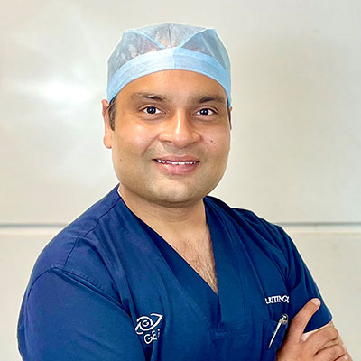
Dr. Anshul Goyal CEO Cataract and Retina Surgeon

Dr. Ritin Goyal Director Cataract and Cornea Surgeon

Dr. Pawan Goyal Chairman Cataract and LASIK Surgeon

Goyal Eye Institute began with a humble beginning in 1989, and has now progressed to provide personalized and inclusive care for entre range of Ophthalmic specialties.

The Centre has highly competent and experienced Ophthalmic Super Specialists to provide best quality care under one roof. Our Specialists form various clinics work closely as a team to provide comprehensive.