Keratoconus: Symptoms, Risk Factors, Stages, & Diagnosis

Hassle free appointment

What is keratoconus?

The thinned cornea and abnormalities on the surface of the cornea are symptoms of keratoconus. The cornea is the front of your eye's transparent outer layer. The cornea's middle layer, its thickest layer, is primarily composed of water and protein collagen. The cornea is made of collagen, which helps maintain its normal, rounded shape and makes it robust and flexible. Having a healthy cornea enables you to see clearly. The cornea thins and bulges into an atypical cone form in keratoconus, impairing vision.

In most cases, keratoconus starts after adolescence and worsens until the mid-30s. It is impossible to forecast whether or how quickly the disease will advance. Both eyes are typically affected by keratoconus, though one is usually more severely affected than the other.

Causes of Keratoconus

What causes keratoconus?

Keratoconus still needs to be fully understood despite decades of research. Although the exact aetiology of keratoconus is unknown, it is thought that a propensity to the condition is present at birth. The loss of corneal collagen is a common finding in keratoconus, and this might be brought on by an imbalance in how corneal cells produce and destroy corneal tissue.

What are the risk factors for keratoconus?

The following factors may boost the chance of acquiring keratoconus:

  • Genetics: Patients are more likely to develop keratoconus if they have Down Syndrome or certain systemic diseases in their family history.
  • Chronic irritation of the eyes: Allergies or other irritants can cause chronic inflammation leading to corneal tissue loss and the development of keratoconus.
  • Glare rubbing: Keratoconus development is linked to persistent eye rubbing. It also increases the likelihood of the disease getting worse.
  • Age: Teenagers are frequently the first to learn they have keratoconus. Younger patients with severe keratoconus are typically more likely to require surgical intervention as the condition worsens.
Risk factors for Keratoconus

What are the symptoms of keratoconus?

Many people with keratoconus are not aware they have the condition. The first sign of the condition is a slight blurring of vision or gradually declining vision that is difficult to repair.

Other keratoconus signs and symptoms include:

  • 1. Lights' halos and glare
  • 2. Night-time vision problems
  • 3. Eye irritability or migraines brought on by eye pain
  • 4. Enhanced sensitivity to intense lighting

How is keratoconus diagnosed?

Your eye care specialist may do the following tests to identify keratoconus in addition to a thorough medical history and eye examination:

  • Corneal topography: This is the most precise method for identifying early keratoconus and tracking its development. A computerised imaging procedure is used to map the corneal curvature.
  • Slit-lamp examination: The outer and middle layers of the cornea can be examined to look for abnormalities.
  • Pachymetry: The cornea's thinnest regions are measured using this test.

How is keratoconus treated?

The course of keratoconus c3r surgery depends on the disease's stage and focuses on vision correction.

Early Stage Treatment for Keratoconus

Early Stages

In the early phases of keratoconus c3r surgery, glasses are used to correct astigmatism and nearsightedness. Glasses can no longer provide patients with a clear vision as keratoconus develops and advances, necessitating the use of a contact lens, typically a hard contact lens.

Intermediate Stages

Cross-linking the corneal collagen is a treatment option for progressive keratoconus. Vitamin B solution is applied to the eye during this one-time office procedure, after which the eye is exposed to ultraviolet light for no more than 30 minutes. New collagen linkages are formed due to the solution, restoring and maintaining some of the cornea's strength and shape.

The procedure can prevent vision from getting worse and, in certain situations, may even enhance vision, but it cannot completely restore the cornea's normal function. To enable the riboflavin to permeate the corneal tissue, the treatment can necessitate the removal of the cornea's thin outer layer (epithelium).

In April 2016, the FDA approved cross-linking as a keratoconus c3r surgery after clinical trials revealed that it prevented or mildly reversed corneal bulging after 3 to 12 months of the surgery.

Intermediate Stage Treatment for Keratoconus
Advanced Stage Treatment for Keratoconus

Advanced Stages

  • Corneal ring: A regular contact lens may become too unpleasant to wear if you have severe keratoconus. Intacs are plastic, implantable C-shaped rings that flatten the cornea's surface to enable better vision. They might also provide a better fit for contact lenses. About 15 minutes are needed for the operation.
  • Corneal transplantation: A donor cornea replaces the patient's injured cornea during a corneal transplant. The procedure for a corneal transplant usually takes one hour and is done as an outpatient. Following the transplant, vision is typically hazy for 3 to 6 months, and medication is required to prevent transplant rejection.

Keratoconus Treatment FAQs

The most effective treatment for keratoconus at the moment is scleral contact lenses. Scleral lenses offer superb comfort and good vision. The most experienced person to fit these specialised lenses in your area will be a local keratoconus contact lens expert.
We cannot completely cure keratoconus, but we can effectively treat it like diabetes and high blood pressure. The preferred method of treatment is frequently scleral contact lenses.
Medical insurance programmes frequently cover the diagnosis and treatment of keratoconus. Contrarily, vision plans typically offer a medically necessary contact lens benefit for using contact lenses to manage keratoconus. Scleral contact lenses are the most typical form of treatment in this situation.
A laser is not used to treat keratoconus. Keratoconus patients cannot have LASIK, PRK, or any other form of refractive surgery. In fact, keratoconus will get worse after refractive surgery
In most cases, keratoconus does not render a person completely blind. However, keratoconus can occasionally impair a person's eyesight to the extent that it interferes with daily activities. With glasses, a patient with advanced keratoconus may have a slight increase in their vision; however, scleral contact lenses may significantly enhance that vision. A corneal transplant may enhance visual acuity if the core corneal scarring is the cause of reduced eyesight. Keratoconus is a highly manageable illness, and most patients notice little change in their daily lives.
There are no natural or homoeopathic remedies for keratoconus.
The majority of keratoconus instances don't require surgery to be properly treated. Glasses can be used as a treatment for mild cases of keratoconus. However, contact lenses will be recommended if the prescription is high. Contact lenses typically treat moderate to severe forms of keratoconus, with scleral lenses having the best success rate.
Patients frequently require a scleral lens to attain their greatest visual acuity following corneal transplant surgery (penetrating keratoplasty).

At Goyal Eye, we have some of the best eye specialists & surgeons

Our specialists

Meet our Team

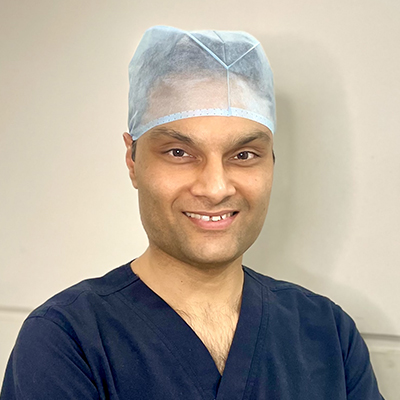
Dr. Anshul Goyal CEO Cataract and Retina Surgeon

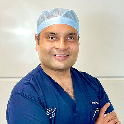
Dr. Ritin Goyal Director Cataract and Cornea Surgeon

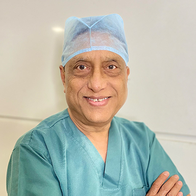
Dr. Pawan Goyal Chairman Cataract and LASIK Surgeon

Goyal Eye Institute began with a humble beginning in 1989, and has now progressed to provide personalized and inclusive care for entre range of Ophthalmic specialties.

The Centre has highly competent and experienced Ophthalmic Super Specialists to provide best quality care under one roof. Our Specialists form various clinics work closely as a team to provide comprehensive.