Understanding Retinal Vein Occlusion: Essential Facts for Eye Health

Hassle free appointment

Retinal Vein Occlusion(RVO)

Retinal Vein Occlusion is a condition in which one or more veins that carry blood away from the retina become blocked or obstructed. This results in the reduced flow of blood to the affected region of the retina. RVO can lead to a number of conditions, including blurry vision, dark spots or floaters, and, in severe cases, complete loss of vision. Individuals at a higher risk of developing RVO include those with a history of diabetes, hypertension, high cholesterol, and heart disease. Smoking and a sedentary lifestyle can also increase the risk of developing RVO.

Untreated RVO can cause serious complications, leading to permanent vision loss and blindness. If you experience sudden disturbances in your vision in any one of the eyes, you should seek medical advice promptly.

Symptoms may include

  • Blurred or distorted vision
  • Partial/ complete loss of vision
  • Sudden onset of floaters
  • Visual field loss
  • A dark or empty spot in your vision

These symptoms might indicate a retinal vein occlusion (RVO). These conditions can cause permanent vision loss or other complications if not treated promptly.
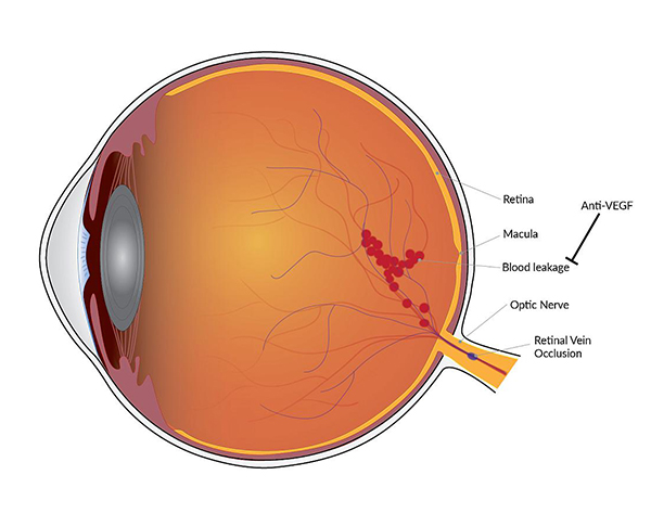
Retinal Vein Occlusion

How does Retinal Vein Occlusion lead to vision loss?

Macular Edema

Macular edema is a medical condition that occurs due to the leakage of blood vessels into the macula (part of the retina), causing it to swell and leading to blurry vision. Macular edema can develop due to a wide range of circumstances. The most prevalent one is diabetic retinopathy, an eye disorder that impairs vision in diabetics. Diabetic macular edema (DME) is the medical term for macular edema that occurs due to diabetic retinopathy.
Macular Edema Retina Condition
Neovascularization Retina Condition

Neovascularization

Neovascularization is a medical condition in which abnormal blood vessels develop in the retina and the light-sensitive tissue at the back of the eye. It can occur due to various eye conditions, including diabetic retinopathy and age-related macular degeneration, and can cause severe vision loss if not treated promptly. Neovascularization frequently manifests in specific types of glaucoma, notably in neovascular glaucoma, which could lead to optic nerve damage and vision loss. The growth of abnormal blood vessels on the iris and/or drainage angle of the eye in this type of glaucoma may increase intraocular pressure, which, if not treated, may result in permanent vision loss. Therefore, it is essential to closely monitor and manage neovascularization in individuals with glaucoma to prevent further harm to their eyesight.

What Experts Say?

Anshul Goyal

Dr. Anshul Goyal

Cataract and Retina Surgeon (CEO)

“Early diagnosis and treatment of retinal vein occlusion can save your eyes from serious complications. Eye injections have made treatment of RVO easy and highly effective. The importance of follow-up with a Retina specialist for at least 6 months cannot be emphasized enough.”

Types of RVO

Central Retinal Vein Occlusion (CRVO)

When a thrombus (blood clot) near the lamina cribrosa blocks the central retinal vein, the condition is called Central Retinal Vein Occlusion (CRVO).

Branch Retinal Vein Occlusion (BRVO)

The second type is called Branch Retinal Vein Occlusion (BRVO), which occurs when a thrombus forms where the retinal artery and vein meet due to atherosclerosis, leading to compression of the retinal vein.

What is the treatment for retinal vein occlusion?

Unfortunately, blocked retinal veins can’t be unblocked again. In cases of swelling or new blood vessel growth(neovascularization), retinal vein occlusion eye injections are typically the go-to treatment option, whereas if there are regions in the retina experiencing significant blood flow deprivation, laser treatment is recommended.

A few retinal vein occlusion treatments include

  • Anti-VEGF Injections: These intra-vitreal injections inhibit the growth of vascular endothelial growth factor(VEGF), which causes macular edema. These injections are a standard treatment for several eye conditions, such as age-related macular degeneration and diabetic retinopathy. Accentrix (Ranibizumab) is considered the most popular anti-VEGF injection. Eyelea is recognized as the most effective option among them. If you’re looking for economic alternatives, go for Avastin (Bevacizumab) & Razumab (Ranibizumab) – a made-in-India product.
  • Corticosteroid Injections: These injections contain drugs that act against inflammatory components which lead to edema.
  • Focal Laser Therapy: It is used to seal off leaking blood vessels and reduce swelling in the affected area of the retina. This can help prevent further damage to the retina. The laser is carefully targeted to the areas of the retina where blood vessels leak, causing damage or swelling. This helps a lot in treating macular edema.
  • Pan-retinal Photocoagulation Therapy: PRP therapy is a medical procedure that uses a laser to treat a wide area of the retina in the eye. The laser creates small burns on the peripheral areas of the retina, which slows down or stops the growth of new blood vessels, preventing further damage to the retina. The procedure can take several sessions, and although it may cause some temporary discomfort or visual disturbance, it is generally safe and effective in reducing the risk of vision loss.

Diagnostics Tests

Are you concerned about your vision? You may be experiencing blurriness or distortion in your eyesight.

If so, it's possible that you're dealing with Retinal Vein Occlusion (RVO).

But don't worry - there are diagnostic tests available to help identify this condition and get you on the path to treatment and recovery. The best doctor for RVO (retinal vein occlusion) is a retina specialist who is specialized in the diagnosis and treatment of retinal diseases, has knowledge and expertise about the latest advancements and treatment options.

Comprehensive eye exam: An eye specialist will perform a detailed examination of the eye, including visual acuity, pupil examination, and a dilated fundus exam to look for signs of RVO.

Retinal vein occlusion (RVO) can occur due to several underlying factors.

  • Atherosclerosis can cause RVO by narrowing the veins with fatty deposits, increasing the risk of blockage.
  • RVO can be caused by a blood clot in the retinal veins due to blood disorders or medications.
  • Hypertension can increase RVO risk by damaging and narrowing the small blood vessels in the retina.
  • In diabetic patients, elevated blood sugar levels can damage retinal blood vessels, increasing the risk of RVO by making them narrowed or leaky.
  • Compression of retinal veins due to eye conditions such as optic disc swelling can cause RVO by narrowing the vessels and reducing blood flow.
  • Blood disorders like polycythemia vera or hyperhomocysteinemia can heighten the RVO risk by thickening the blood and increasing the likelihood of blood clots and blockages.
  • Inflammatory conditions like giant cell arteritis or lupus can cause RVO by damaging blood vessels and increasing the risk of blockages.
  • Medications like oral contraceptives or hormone replacement therapy can heighten the RVO risk by increasing blood thickness and the likelihood of blood clots.

Prevention Of Retinal Vein Occlusion

Retinal vein occlusion and cardiovascular diseases have similar reasons to occur. Here are a few things you can do to prevent yourself from cardiovascular diseases as well as RVO.

  • Treatment of high blood pressure
  • Reducing the bad cholesterol level
  • Quitting smoking
  • Control your eating habits if you’re diabetic
  • Weight loss if you are overweight.
Prevention Of Retinal Vein Occlusion

FAQs

Retinal vein occlusion can generally occur in one eye.
Retinal vein occlusion occurs more frequently in older adults or those with underlying medical conditions like diabetes or high cholesterol.
To diagnose retinal vein occlusion, an ophthalmologist can perform a comprehensive eye exam that includes a dilated eye exam, a visual acuity test, and imaging tests such as optical coherence tomography (OCT) or fluorescein angiography.
Risk factors for retinal vein occlusion include high blood pressure, diabetes, smoking, high cholesterol, and a history of blood clots.
No, Retinal vein occlusion can’t be inherited.
Retinal vein occlusion can recur after treatment, so regular eye exams and close monitoring are crucial to detect any changes in your vision.

At Goyal Eye, we have some of the best eye specialists & surgeons

Our specialists

Meet our Team

Dr. Anshul Goyal CEO Cataract and Retina Surgeon

Dr. Ritin Goyal Director Cataract and Cornea Surgeon

Dr. Pawan Goyal Chairman Cataract and LASIK Surgeon

Goyal Eye Institute began with a humble beginning in 1989, and has now progressed to provide personalized and inclusive care for entre range of Ophthalmic specialties.

The Centre has highly competent and experienced Ophthalmic Super Specialists to provide best quality care under one roof. Our Specialists form various clinics work closely as a team to provide comprehensive.