Symptoms of CRAO
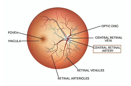
The main symptom of CRAO is a sudden and painless loss of vision in one eye, resulting in a complete or near-complete visual loss. The onset is typically rapid and may occur within seconds or minutes. Some individuals may experience a curtain-like effect or a darkening of vision before the complete loss.
Causes of Central retinal artery occlusion (CRAO)
CRAO is most commonly caused by a blood clot or embolus that blocks the central retinal artery. The embolus typically originates from the carotid artery or the heart and travels through the bloodstream, eventually lodging in the smaller vessels of the eye. Other less common causes include inflammation, compression of the artery, or vascular diseases affecting the blood vessels in the eye.
Certain health conditionings such as advanced age, hypertension (high blood pressure), diabetes, smoking, hyperlipidemia (high cholesterol), and a history of previous transient ischemic attacks (TIAs) or stroke can cause CRAO.
Treatment of CRAO
Central retinal artery occlusion treatment aims to restore blood flow in the retina and prevent further damage in vision loss. Although there is no treatment if the vision gets completely lost, a certain vision level can still be regained. It also depends on the issue's duration and the damage level in the retina. These are the best treatments to save the eyes from complete blindness:
- Ocular Massage: Digital massage of the globe of the affected eye may be performed to dislodge the embolus and improve blood flow. This technique should only be attempted by a qualified healthcare professional or ophthalmologist.
- Inhalation of Carbon Dioxide (Carbogen Therapy): Breathing in a mixture of carbon dioxide and oxygen, known as carbogen therapy, can help dilate blood vessels and improve oxygenation to the retina. This treatment is typically administered in a hospital or clinical setting.
- Medications:
a. Acetazolamide: It helps reduce fluid buildup in the eye and may be prescribed to lower intraocular pressure and improve blood flow to the retina.
b. Hyperbaric Oxygen Therapy (HBOT): HBOT involves breathing pure oxygen in a pressurized chamber, which can enhance oxygen delivery to tissues, including the retina. - Thrombolysis: In certain instances, thrombolytic therapy may be considered to dissolve the blood clot causing the occlusion. Thrombolytic drugs, such as tissue plasminogen activator (TPA), can help restore blood flow.
- Intra-arterial Thrombolysis and Mechanical Thrombectomy: In more severe cases, when a visible clot causes the occlusion, specialized procedures like intra-arterial thrombolysis or mechanical thrombectomy may be attempted. These procedures remove the clot directly using catheters or devices, typically performed by interventional ophthalmologists or vascular specialists.
Diagnosis of CRAO
Central Retinal Artery Occlusion diagnosis involves a comprehensive evaluation of the patient's medical history, symptoms and a thorough eye examination. These are the common diagnostic procedures:
- Visual Acuity Testing: The patient's visual acuity is used as an eye chart to determine the extent of vision loss.
- Ophthalmoscopy: This examination involves using a specialized instrument called an ophthalmoscope to examine the back of the eye, including the retina and optic nerve.
- Fluorescein Angiography: This test involves injecting a fluorescent dye into a vein in the patient's arm. As the dye circulates through the blood vessels in the eye, photographs are taken to evaluate the blood flow and identify any blockages or abnormalities. Fluorescein angiography helps visualize the occluded central retinal artery and its branches.
- Optical Coherence Tomography (OCT): This non-invasive imaging test uses light waves to capture detailed cross-sectional images of the retina. OCT can provide information about the thickness and integrity of retinal layers, allowing doctors to assess the extent of retinal damage.
- Carotid Ultrasound/Doppler Imaging: An ultrasound or Doppler imaging may be performed to assess the health of the carotid arteries, which supply blood to the head and neck. These tests can help detect any blockages or narrowing of the arteries that could contribute to emboli formation.
What are the complications?
The growth of abnormal blood vessels in the iris, retina, or angle are common complications. It can result in further vision loss or pain in the affected area of the eye. These irregular blood vessels cannot Ibe found in patients treated with tPA or hyperbaric oxygen. Therefore, these are treated with anti-VEGF agents or surgery if they increase to vitreous hemorrhage or uncontrolled glaucoma.
Treatment of Complications
Although the visual loss is quite severe and entirely depends on the amount of retina damage. However, with the best treatments, such as cilioretinal artery and visual acuity, 20/50 or more recovery happens in over 80% of eyes.
Prevention from CRAO
CRAO is generally connected to diabetes or heart problems, but they don't directly cause that. Yet, a safe and preventive step is to take care of your heart by doing these things:
- Maintain a healthy weight
- Consuming a healthy diet
- Daily exercises
- Stop smoking and alcohol
- Keep blood sugar levels at normal in case of diabetes
FAQs for CRAO
At Goyal Eye, we have some of the best eye specialists & surgeons
Meet our Team

Dr. Anshul Goyal CEO Cataract and Retina Surgeon

Dr. Ritin Goyal Director Cataract and Cornea Surgeon

Dr. Pawan Goyal Chairman Cataract and LASIK Surgeon

Goyal Eye Institute began with a humble beginning in 1989, and has now progressed to provide personalized and inclusive care for entre range of Ophthalmic specialties.
The Centre has highly competent and experienced Ophthalmic Super Specialists to provide best quality care under one roof. Our Specialists form various clinics work closely as a team to provide comprehensive.
Eye Issues
- Red Eyes
- Dry Eyes
- Blurry Vision
- Keratoconus
- Diabetic Retinopathy
- Strabismus
- Retinal Vein Occlusion
- Retina Pigmentosa
- Retinal Tear
- Floaters
- Vitreous Haemorrhage (VH)
- Central Retinal Artery Occlusion (CRAO)
- Central Serous Chorioretinopathy (CSCR)
- Cystoid Macular Edema (CME)
- Retinopathy of Prematurity (ROP)
- Age-Related Macular Degeneration (ARMD)
© Copyright Goyal Eye, All Rights Reserved - 2026
